More than just cholesterol
Artery plaques, heart attacks more complicated than that
9/1/2014
The Blade
Buy This Image

Robert Lee
This is one in a series of articles written by research students in The University of Toledo College of Medicine’s Biomedical graduate program exploring basic issues of human health.
Most people have seen the commercials with the simple animation of cholesterol floating through the blood and then sticking to the wall of the blood vessel. It piles up, forming something called a plaque that continues to grow until, eventually, it fills up the whole artery, blocking blood flow and causing a heart attack. But is it really as simple as that?
The process of plaque buildup, a disease called atherosclerosis, is almost always associated with high cholesterol levels, but the plaque itself is made up of more than just cholesterol.
Atherosclerosis is an inflammatory disease. Cells that act as “protective police” within the body cause redness and swelling, called inflammation, around a scratch. These same protective cells are attracted to the artery wall when cholesterol starts to build up, because this also is recognized as an injury.
The immune system is made of cells that can fight off infection from bacteria by recognizing certain parts of bacteria as foreign to the human body. In the same way, cholesterol that accumulates in the artery wall is recognized as foreign. One type of immune cell, called a macrophage, is one of the first cells “on the scene” when cholesterol accumulation occurs.
It has been more than a century since macrophages were discovered in atherosclerosis, but researchers are just now starting to truly appreciate the important role they play in the development of dangerous plaques that can lead to cardiac events. These plaques begin to develop very early on in life, and have been observed in teenagers and even younger children. However, the risk of dying from a heart attack at a young age is extremely low.
So, what happens to these plaques over a lifetime to make them more deadly?
One of the biggest developments in the last few decades in the field of atherosclerosis is the discovery that inside the plaque, many of the macrophages are dying. Cell death is a normal process in the body, and at the end of their lifespan many cells undergo a controlled form of death called apoptosis.
Dying cells are recognized by other healthy cells and are removed from the tissue. This process of clearing out dead cells is extremely important. Macrophages that die without being cleaned up can eventually start to become "necrotic," another name for becoming rotten, which will cause inflammation.
Imagine these dying macrophages as dying animals, and the cells that clean them up as vultures. If there were no vultures to eat and clean up the bodies of animals that have died, the bodies would become rotten, start to stink and attract loads of flies and other bugs, and more and more carcasses would be found. Similarly, there is now evidence that in dangerous plaques, the cells that act as vultures are not able to do their job properly, and the corpses of macrophages build up in the center of the plaque. This is called a "macrophage graveyard."
So why are we worried about this "macrophage graveyard" and how is it linked to risk of heart attack?
Cells are filled with fluids and other molecules that help them function properly. When a macrophage dies and becomes rotten, it can essentially explode and release all of its contents into the plaque. These molecules that are normally found inside the cell, once released into the surrounding area, can cause the plaque to become more likely to rupture into the artery. A plaque breaking open is what causes the blood flow in the artery to be cut off and results in a heart attack.
A major focus of our research at the University of Toledo during the last few years has been to investigate how to prevent macrophages from dying inside the plaque. Currently, we are studying how we can turn on proteins on the surface of macrophages, called receptors, to try to stop macrophage death.
We found that when we treated dying macrophages with a drug to turn on one of these receptors, we could reduce the number of macrophages that died. When activated, the receptors are able to transmit anti-death signals inside the macrophage to keep the cell alive.
Our next goal is to find a way to reproduce this anti-death effect inside the plaque. We might be able to prevent the buildup of rotting cells and reduce the risk of the plaque from rupturing and causing heart attacks by finding new discoveries within this important area of biomedical research.
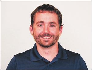
Robert Lee is a PhD graduate student at the University of Toledo College of Medicine and Life Sciences Biomedical Science Program, the former Medical College of Ohio. For more information, email Robert.Lee5@rockets.utoledo.edu or go to utoledo.edu/med/grad/biomedical.