Body of research: Cadavers become teachers in medical school classrooms
9/18/2010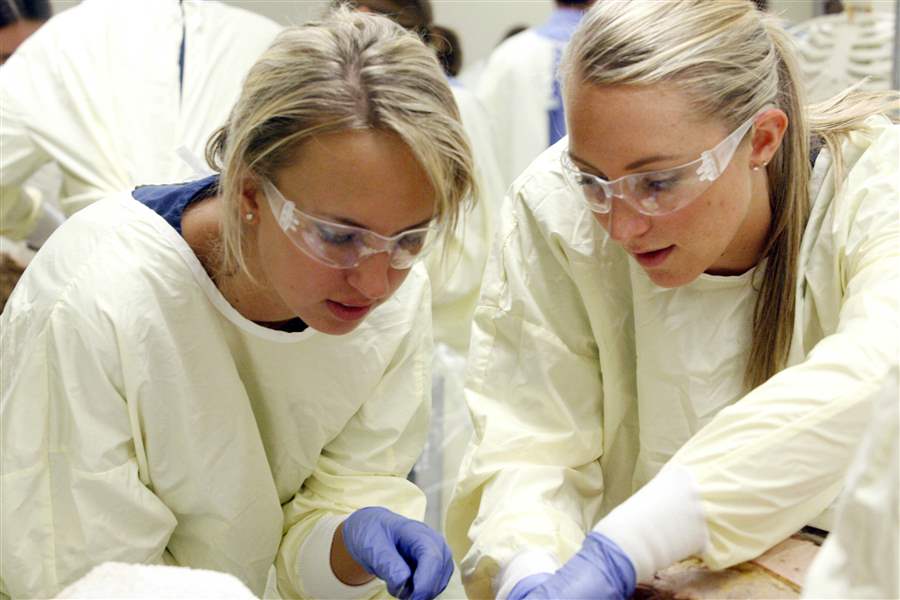
Ashley Sheskey, left, and Natalie Kerestes work together in the anatomy lab.
The Blade/Amy E. Voigt
Buy This Image

Ashley Sheskey, left, and Natalie Kerestes work together in the anatomy lab.
Resting peacefully flat on his back, the elderly first patient of student Jenna Yeager yields few clues about his background or maladies. She knows only that he is 87 years old and, at some point, developed coronary artery disease.
The man cannot cooperate in the medical investigation that Ms. Yeager and her fellow physician assistants-in-training are about to undertake because he is deceased. His is among six cadavers being dissected on this day by Ms. Yeager and her classmates in an anatomy lab at the University of Toledo Medical School, the former Medical College of Ohio. Students do not know names of cadavers, only their age and cause of death.
To some people who talk loftily of donating their bodies to medicine, this room in the Block Health Science Building would come as something of a disappointment. It has a pungent odor and is filled not with senior researchers making scientific breakthroughs but three dozen first-year physician's assistants-in-training sawing and cutting their way through the human chest cavity as they make mistakes and gain insights.
"I was really excited about doing this," says Ms. Yeager. "It supplements classroom lectures. The best way to learn is to see something. Once I see it, it ties the lecture together."

Mark Hankin, left, instructs students Robert Bell and Hope Dundas.
Mark Hankin, professor of anatomy and director of UT's anatomical donations program, tells students that the cadavers are their first patients. And he is convinced that what takes place here is as important as activity in any research lab.
Each student trained here has the potential to affect thousands of patients over a medical career, Mr. Hankin points out. "Anatomy is the basis of medicine," he says of the field concerned with the structure and function of the human body.
Interest is growing
Painted in bold black on a rear wall of the room is a famous inscription borrowed from an early Paris anatomy-teaching theater: "Here is the Place where Death Enjoys Helping Life."
A variation of that inscription is etched on a stone in Woodlawn Cemetery that marks the common grave where cremated remains are buried when students are finished with the bodies.
About 4,000 people have agreed to donate their bodies to UT, and interest is growing.
Diane Durliat, coordinator of the anatomical donation program, attributes the upsurge partly to people seeking to avoid funeral expenses in a poor economy.
This year, 275 people requested information packets regarding donating. That is more than double the number of 2008. If past patterns hold, 80 percent of those who make inquiries will pay the $100 fee and sign papers turning over their bodies to the medical school when they die, the coordinator says. Donors must be signed up in advance, and family members are not permitted to make donations after death, she adds.
The number of bodies has doubled to 200 a year since Ms. Durliat became coordinator seven years ago. The school receives no bodies from public institutions that deal with people who are indigent.
Besides financial considerations, donors are motivated by the desire to advance patient care, she says.
Sometimes, donors who don't fully understand the program ask that their families receive reports on "what we find," the coordinator says. Program personnel gently explain that nearly all cadavers are used to teach students in various fields of medicine to recognize body parts and systems and that "we're not studying the cause of death," says Ms. Durliat.
So far, that hasn't led anyone to back out, she adds.
Eureka moments
On a blazingly hot recent afternoon, Ms. Yeager and 35 classmates filed into the anatomy lab for a two-hour lesson that will focus on the aorta, esophagus, and other parts that remain in the chest after removal of the heart and lungs. Extracted in prior sessions, the two organs sit in zippered plastic bags near bodies.
Clad in yellow smocks, the students are gathered in groups of five or six over individual cadavers. Students, guided by a workbook called Grant's Dissector, start with the back and then move to the chest, upper limbs, abdomen, pelvis, lower limbs, neck, and head.
Ms. Yeager's group, in a class prior to this, their fourth session, had a eureka moment. They found telltale signs that the man whose cadaver they are dissecting — dubbed "Fred" by one group member — underwent coronary bypass surgery.
Discovering a physical anomaly as the group works through today's lesson, one student incorrectly concludes that this was the result of the bypass surgery. "It's not from the bypass," explains Richard Lane, professor of anatomy and co-director of the anatomical donation program. "He was born like that."
"Maybe he got jumbled up too much when he was a baby — His mom was a runner," jokes one group member in the kind of good-natured, but not disrespectful, banter that is commonplace in the anatomy lab.
Dilemmas
More seriously, students explain that the class has helped them to understand the person-to-person differences in how individual body parts and systems are positioned and that there is little variability in how they are connected to each other.
Cloths cover the faces of each cadaver, although students are not required to do so.
At another dissection station, students working with the body of a woman who died of gangrene and dementia at 86 debate the appropriateness of concealing the face.
Thirty-seven-year-old Hope Dundas says she would have preferred that the cloth be left off. "It connects me to the person," explains the Ann Arbor native. "It lets me see that it's not just parts."
Robert Bell, who acknowledges that the initial sight of a human cadaver made him uneasy, appreciates the covering. It is easier to complete dissection exercises if the person's humanity isn't always top-of-mind, explains the 22-year-old from Cleveland.
"The majority of people said to cover it, so it's no big deal," shrugs Ms. Dundas.
The group has not assigned a name to the cadaver, as is common in medical school anatomy labs.
"To me, it's kind of disrespectful," says Ms. Dundas. "This was a person in real life. She had an individual identity."
Excitement
Group members admit to initial queasiness. "You get a little bit of butterflies each time you come to class," acknowledges Kellie Schoenlein, 23, of Maria Stein, Ohio, near Lima. "But you're here to learn ..."
Andrew St. Pierre of Monroe is comforted by the knowledge that: "These people made a choice to let you learn from them."
Students agree that they gain valuable knowledge from the class. "You get to mix this up with what you've learned in the books," says Joe Rose, of Whitehouse. "This is something you're going to recall so that 10 years down the road when you have a patient with this, you recognize it."
Mr. Hankin, the program director, says some students are initially anxious about working with cadavers, but he was unaware of any who dropped out of the class.
The experiences can be especially troubling for a student who has recently lost a grandparent or other loved one, he says.
"When I lost my mother, it got to me when I saw people who had a similar disease or the wasting that she had," he adds.
When students experience serious difficulty, counseling is available from faculty members and professional counselors, he says
For the most part, however, Mr. Hankin says, "Students approach anatomy with excitement ... People are really interested in their bodies. Everybody wants to see what's inside it. These students are no different."
Advanced training
With the availability of interactive computer products for teaching dissection, there is debate among medical educators about the continued use of human cadavers. At least one medical school, in the United Kingdom, has gone completely digital.
But anatomy lab instructors and staff at UT plan no such move.
"There is a sense that comes from touching the body structures and muscles and nerves" that students wouldn't get on a computer, says lab supervisor Beth Dalzell, who also embalms bodies. "Since we get so many different donors, everybody is different. Students get to see a whole range of things."
With advances such as haptics technology, it is possible that students will one day be able to gain a sense of touch with computerized dissection programs, Mr. Hankin acknowledges. But such technology is expensive, and widespread use is decades away, he adds.
"Interacting with a real body that was a living person teaches students something very different than they can get with a computer," he continues. "And would you want your doctor to train only on a computer before he operates on you? Most people would say no."
Contact Gary Pakulski at:
gpakulski@theblade.com
or 419-724-6082.