Zebrafish help in the fight against cancer
4/4/2016
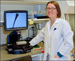
Kari Lavik is a postdoctoral fellow in the department of biochemistry and cancer biology at the University of Toledo college of medicine and life sciences.
Nearly everyone has been touched by cancer.
According to the American Cancer Society, 14.5 million Americans were living with or in remission from cancer in 2014. Whether cancer is found within you, a friend, a loved one, or an icon like the late David Bowie, a cancer diagnosis always reminds us of our mortality and how quickly our lives can change.
Finding a cure for cancer is a daunting task. Cancer is a complicated disease, with treatment depending on the organ and person affected. Cancer is constantly growing and changing. It is a snake with many heads. If you chop off one head, or treat one aspect of the disease, a new head may grow in its place. Cancer cells are constantly evolving and learning how to survive. To conquer this beast we need to attack multiple heads at once.
To speed up the investigative process for new treatment options, scientists at the University of Toledo’s college of medicine and life sciences, the former Medical College of Ohio, have begun using a zebrafish model as a new way of screening anti-cancer drugs.
Current cancer treatment strategies include surgery, radiation, and a short laundry list of drugs that doctors can choose from depending on each patient’s diagnosis. Some drugs prevent cancer cells from growing, while others prevent cells from moving and still others can kill cancer cells in their tracks. The problem is that over time many cancer cells learn how to resist the limited list of treatments currently available. Medical researchers are always on the hunt for new and better ways to fight cancer in its many manifestations.
Developing therapies for human use is exceedingly difficult. Drug candidates are subjected to rigorous testing to maximize treatment success and patient safety. The magic balance that is so difficult to achieve is to maximize killing or inhibiting growth of cancer cells while minimizing harm to normal cells within the patient. Drug screening often starts in cells that are cultured in dishes. Scientists can grow human cancer cells in dishes to carefully study how they behave. We can then assess how the cancer cells respond to each drug that they are treated with. Do the cells stop growing? Do they change how they move? Do they die?
At UT, researchers have been diligently studying a series of new anti-cancer drug candidates, which we hope will some day improve patient survival. For example, glioblastoma multiforme is a devastating cancer of the brain because these cancer cells invade rapidly, making surgical removal and further treatment difficult. Our research is focused on investigating a series of drugs that prevent glioblastoma cancer cell movement. By blocking the cancer cells from moving, treatment and surgical removal can be vastly improved.
Similarly, our colleagues within the UT Department of Biochemistry and Cancer Biology have developed a different drug that can actually kill glioblastoma cells. This drug triggers the glioblastoma cells to fill up with fluid-filled bubbles called vacuoles. Over time, the cancer cells become overwhelmed by vacuoles and die.
Both of these drug candidates have shown great promise; however, before they can be approved for clinical trials in humans, they must first undergo rigorous screening in animal models. Historically, such screens have been performed in mouse and rat models. Unfortunately, these types of studies are laborious, expensive and take years to complete. The zebrafish model provides another option.
Zebrafish are small minnows, native to India and the surrounding region, which have been gaining popularity among scientists for biomedical research. These small fish are ideal for biomedical studies, because the young fish are transparent, allowing researchers to easily observe organ development and function. Furthermore, cancer cells that are stained with special dyes can be implanted into young zebrafish in order to visually track how cancer cells grow and spread.
Currently, we are introducing specifically labeled glioblastoma cells into the yolk sac of young zebrafish. These cancer cells glow under specific wavelengths of light so that we can directly observe them growing and spreading throughout the fish. We have now identified a range of concentrations of our investigative anti-cancer drugs that do not harm the fish and should stop the implanted cancer cells from moving, according to cell culture studies.
In future studies, we plan to treat the cancer cells in the young fish with our drugs to both prevent cancer cell invasion and promote cancer cell death. By using the zebrafish model, we can compress years of work into a few months and spend a fraction of the cost to more quickly identify safe and efficient cancer treatments, allowing us to speed up the process of saving lives.
Kari Lavik is a postdoctoral fellow in the department of biochemistry and cancer biology at the University of Toledo college of medicine and life sciences. She is doing her research in the laboratory of Dr. Kathryn Eisenmann. For more information, contact Kari.Lavik@rockets.utoledo.edu or go to utoledo.edu/med/grad/biomedical.